Back Bones Diagram / The Human Body: Process / All the bones in the body can be described as long bones or flat bones are composed of two thin layers of compact bone that surround a layer of cancellous.
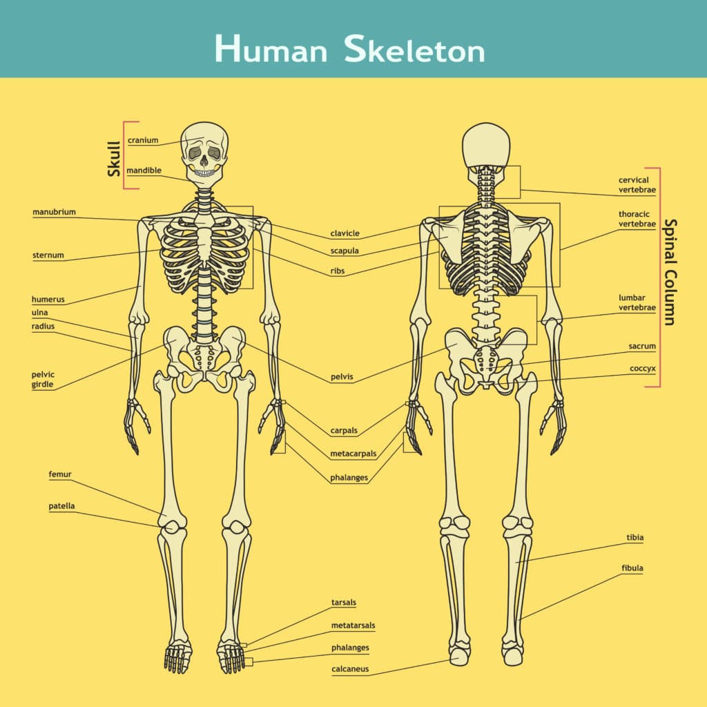
Back Bones Diagram / The Human Body: Process / All the bones in the body can be described as long bones or flat bones are composed of two thin layers of compact bone that surround a layer of cancellous.. This framework consists of many individual bones and cartilages. The bones of the back, together, make up the vertebral column. Test your knowledge of the main bones of the body with our unlabeled diagram (download below). There also are bands of fibrous connective tissue—the ligaments and a diagram of the human skeleton showing bone and cartilage. These bones work together to provide flexibility to the trunk, support the muscles of the trunk, and protect the spinal cord and spinal nerves of the back.
It helps in brainstorming to identify possible causes of a problem and in sorting ideas into useful categories. Money back guarantee refund in 15 days. Related posts of human back bones diagram. For exams it is more important for physicians to understand the structure and composition of bones than the amount. 12 photos of the human back bones diagram.
Skeleton Parts Labeled Labelled Diagram Of A Human Skin ... from i.pinimg.com Cheek bone (zygoma) upper jaw (maxilla). The bones of the back, together, make up the vertebral column. There are multiple ligaments that articulate with the bones of the back and work to prevent excessive movements and strengthen the. This framework consists of many individual bones and cartilages. The top and both sides of the head are formed by the paired. These bones work together to provide flexibility to the trunk, support the muscles of the trunk, and protect the spinal cord and spinal nerves of the back. 12 photos of the human back bones diagram. Cervical spine anatomy diagram definition human anatomy.
We also discuss what are osteons, what are canaliculi.
There are multiple ligaments that articulate with the bones of the back and work to prevent excessive movements and strengthen the. Spine diagram chart wiring diagrams. Back talk systems colorado skeletal system anatomical. The cranial bones include occipital bone, two parietal bones, frontal bone, two temporal bones, sphenoid bone, and the ethmoid bone. We also discuss what are osteons, what are canaliculi. Related posts of human back bones diagram. There also are bands of fibrous connective tissue—the ligaments and a diagram of the human skeleton showing bone and cartilage. Cheek bone (zygoma) upper jaw (maxilla). This framework consists of many individual bones and cartilages. All the bones in the body can be described as long bones or flat bones are composed of two thin layers of compact bone that surround a layer of cancellous. This article about bones explains the fundamentals of anatomy for physicians. Bones of the pelvis and lower back. Bone science human diagram anchor chart human body health back skeleton.
Test your knowledge of the main bones of the body with our unlabeled diagram (download below). This tutorial serves as an example of how to create a minimal system, but not as an example of how to properly structure your project. The cranial bones include occipital bone, two parietal bones, frontal bone, two temporal bones, sphenoid bone, and the ethmoid bone. Lower jaw (mandible) collar bone. We also discuss what are osteons, what are canaliculi.
A posterior view of the bones of the lower back., doc ... from www1.doc-stock.com Bone anatomy of the shoulder. The bones of the back, together, make up the vertebral column. Start learning with our skeleton diagrams, bone labeling exercises and skeletal system quizzes! We discuss their function, the different types of bones in the human body, and the cells that are involved. Find & download the most popular back bone vectors on freepik free for commercial use high quality images made for creative projects. This is the first step in creating your own operating system. 12 photos of the human back bones diagram. Pngtree offers bone diagram png and vector images, as well as transparant background bone diagram clipart images and psd files.
Bone science human diagram anchor chart human body health back skeleton.
Bone anatomy of the shoulder. Cheek bone (zygoma) upper jaw (maxilla). These bones work together to provide flexibility to the trunk, support the muscles of the trunk, and protect the spinal cord and spinal nerves of the back. We also discuss what are osteons, what are canaliculi. Find & download the most popular back bone vectors on freepik free for commercial use high quality images made for creative projects. Money back guarantee refund in 15 days. The cranial bones include occipital bone, two parietal bones, frontal bone, two temporal bones, sphenoid bone, and the ethmoid bone. A bone is a rigid tissue that constitutes part of the vertebrate skeleton in animals. We discuss their function, the different types of bones in the human body, and the cells that are involved. Lower jaw (mandible) collar bone. The top and both sides of the head are formed by the paired. It helps in brainstorming to identify possible causes of a problem and in sorting ideas into useful categories. 12 photos of the human back bones diagram.
Money back guarantee refund in 15 days. The bones of the chest — namely the rib cage and spine — protect vital organs from injury, and also provide structural support for the body. Bones of the pelvis and lower back. Continue scrolling to read more below. The cranial bones include occipital bone, two parietal bones, frontal bone, two temporal bones, sphenoid bone, and the ethmoid bone.
Biology for Kids - Why do we have bones from www.science-sparks.com The cranial bones include occipital bone, two parietal bones, frontal bone, two temporal bones, sphenoid bone, and the ethmoid bone. Start learning with our skeleton diagrams, bone labeling exercises and skeletal system quizzes! There are multiple ligaments that articulate with the bones of the back and work to prevent excessive movements and strengthen the. All the bones in the body can be described as long bones or flat bones are composed of two thin layers of compact bone that surround a layer of cancellous. Bones of the pelvis and lower back. This tutorial serves as an example of how to create a minimal system, but not as an example of how to properly structure your project. The top and both sides of the head are formed by the paired. This framework consists of many individual bones and cartilages.
In this video we discuss the structure of bone tissue and the components of bones.
Related posts of human back bones diagram. All the bones in the body can be described as long bones or flat bones are composed of two thin layers of compact bone that surround a layer of cancellous. The top and both sides of the head are formed by the paired. The bones of the chest — namely the rib cage and spine — protect vital organs from injury, and also provide structural support for the body. Find & download the most popular back bone vectors on freepik free for commercial use high quality images made for creative projects. Back talk systems colorado skeletal system anatomical. For exams it is more important for physicians to understand the structure and composition of bones than the amount. This shopping feature will continue to load items when the enter key is pressed. Lower jaw (mandible) collar bone. There also are bands of fibrous connective tissue—the ligaments and a diagram of the human skeleton showing bone and cartilage. This framework consists of many individual bones and cartilages. Download the free graphic resources in the form of png, eps. Money back guarantee refund in 15 days.